ALERT
ALERT For support with UOM FDM, please raise a ServiceNow ticket https://uomservicehub.service-now.com/esc?id=sc_cat_item&sys_id=7b8de37e1b1f201cef32a68b274bcbb2.
(hide this warning on this page)
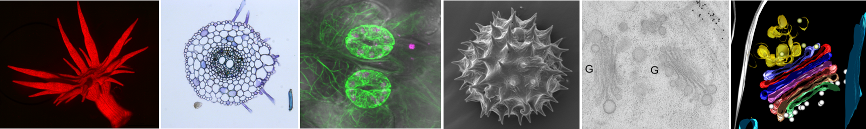
The BioSciences Microscopy Unit (BMU) offers a range of imaging techniques to help life science researchers magnify and investigate the dynamics, structure and function of biological samples.
The range of imaging and preparation techniques of the BMU include light, fluorescence, confocal and electron microscopy. Both transmission and scanning electron microscopy (TEM & SEM) are available with electron tomography and entry-level cryoEM on the TEM, and elemental analysis (EDS) on the SEM.
We work with academic and commercial researchers to help investigate different biological samples at organism, tissue, cell, organelle and molecular levels.
We can help with:
• Instrument training
• Microscopy applications training
• Collaboration in optical and electron microscopy techniques for research projects
| Allison van de Meene (PhD) |
| Core Manager, BioSciences Microscopy Unit |
| 03 8344 9828 |
| biosciences-microscopy@unimelb.edu.au |
| Hours | Location |
|
Monday - Friday 9 am - 5 pm |
Biosciences 2 |
| Name | Role | Phone | Location | |
|---|---|---|---|---|
| General Enquiries |
03 8344 9828
|
biosciences-microscopy@unimelb.edu.au
|
BioSciences 2, Parkville, The University of Melbourne
|
| ► Confocal Laser Microscopy (3) | ||||||||||||
|
||||||||||||
| ► Multi-instrument rooms (2) | ||||||||||||
|
||||||||||||